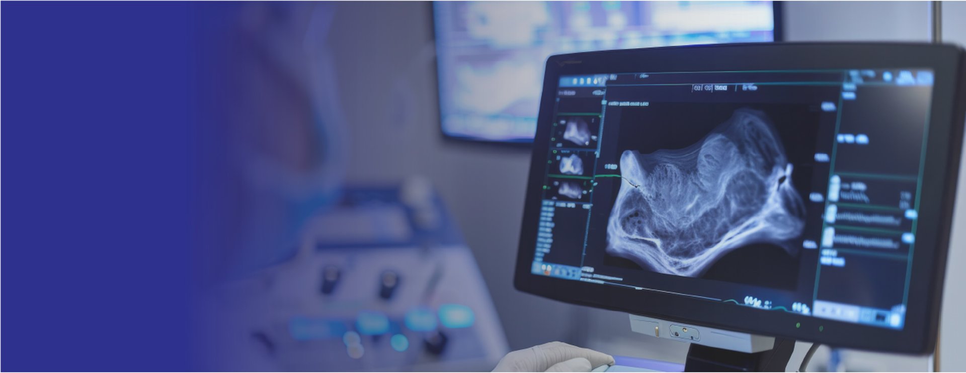
Ultrasound in Women’s Health
Safe, Gentle Imaging – See What Matters Most
Ultrasound uses harmless sound waves to create clear pictures of what’s happening inside your body. It’s safe (no radiation), painless, and a valuable tool for both pregnancy care and gynaecological health.
Ultrasound in Gynaecology
What does it check?
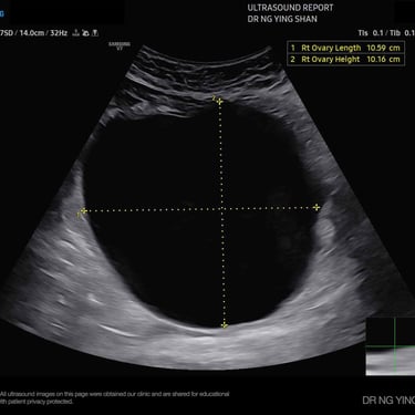
The size, shape, and lining of the womb(uterus)
The ovaries and developing eggs (follicles)
Other structures in the pelvis, including any lumps or growths

Common conditions it can detect:
Fibroids (non-cancerous growths in the womb)
Ovarian cysts (fluid-filled sacs in the ovary)
PCOS (polycystic ovary syndrome)
Signs of endometriosis or adenomyosis
Possible causes of heavy or irregular periods
If you’re experiencing symptoms like unusual bleeding, pelvic pain, irregular periods, or have concerns about fertility, a gynaecological ultrasound can help find answers quickly and safely. It uses sound waves to create clear images of your reproductive organs


This scan is usually quick, painless, and provides important information to
guide the right treatment for you.
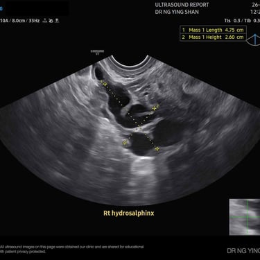
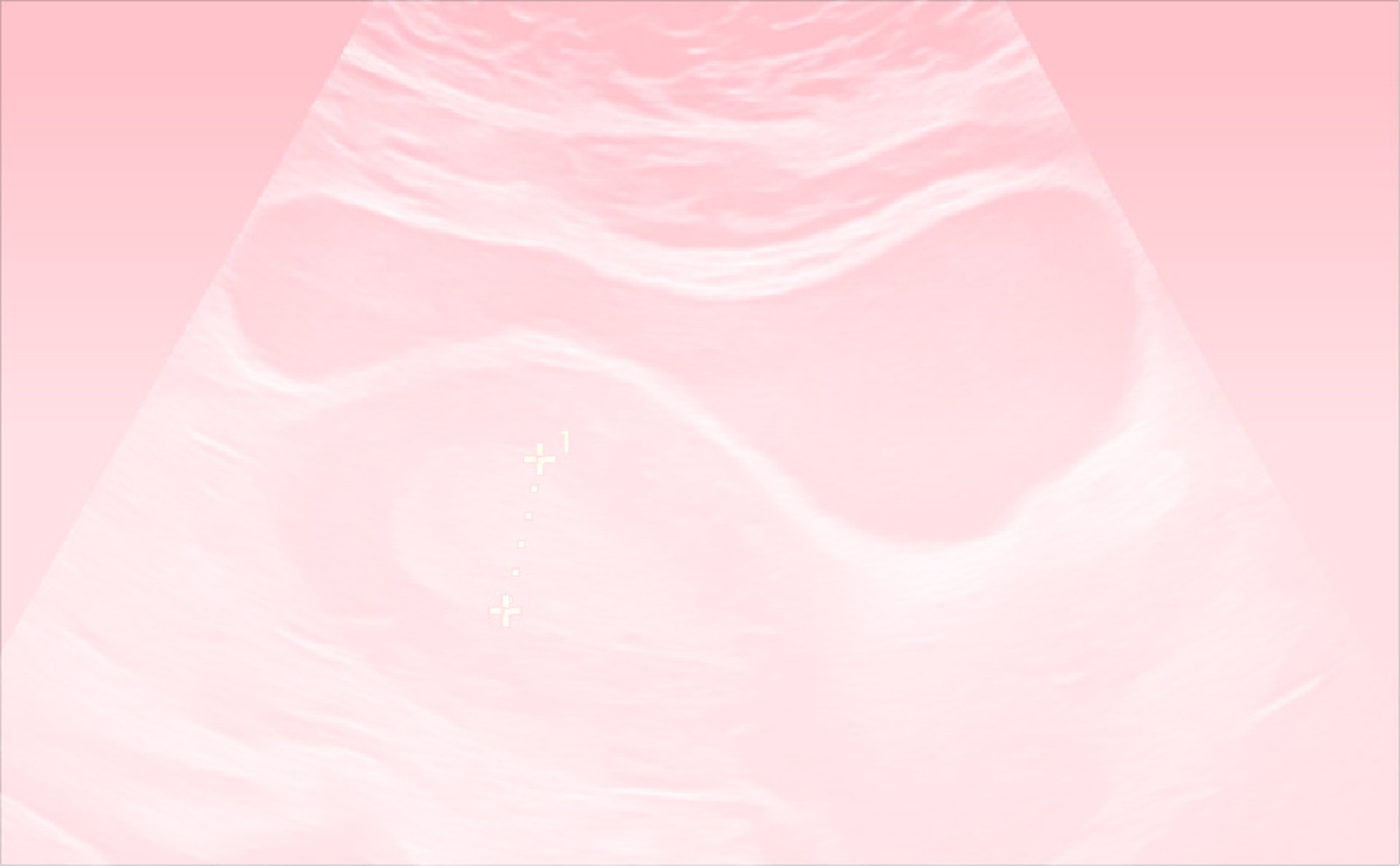
hydrosalphinx
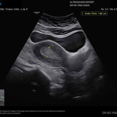
Thickened ET
Ovarian Cyst
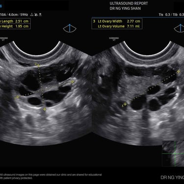
Polycystic Ovary
The Two Main Types of
Gynaecological Ultrasound
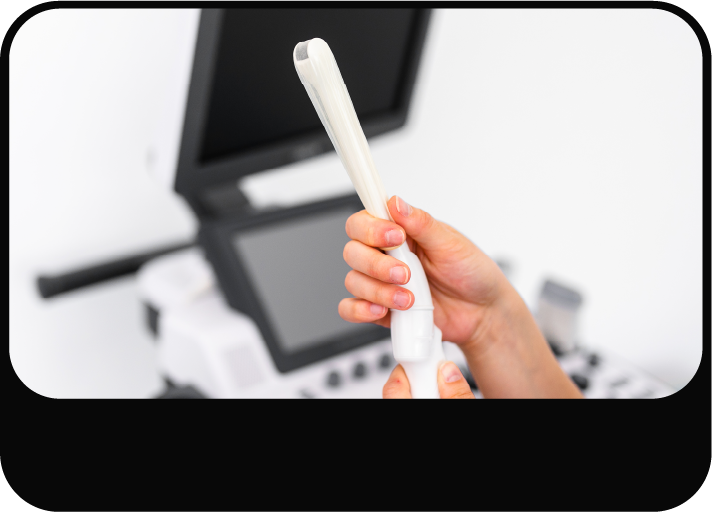
Gel is applied to your lower tummy; a probe glides over the skin. It’s comfortable and used when a broader view is needed or transvaginal isn’t suitable.
A slim, covered probe is gently inserted into the vagina (like a tampon).
It gives sharper, closer images – especially helpful in early pregnancy or detailed gynaecology checks.
It gives clearer pictures on the scan, so doctors can see the organs in the lower belly area more clearly and understand what might be wrong
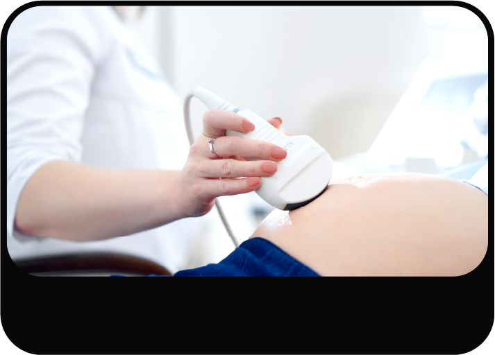


Abdominal Ultrasound
Transvaginal Ultrasound
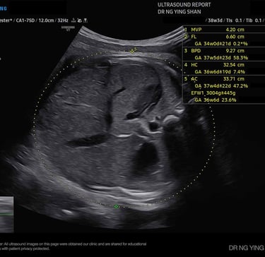
Ultrasound in Pregnancy
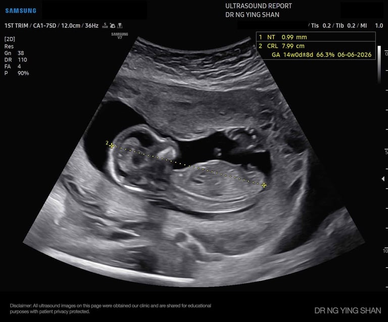
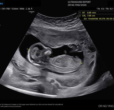
Ultrasound is performed in pregnancy to confirm the pregnancy’s location and viability, determine the number of fetuses, monitor the baby’s growth and development, check the placenta and amniotic fluid, and screen for any potential issues.
6-10
weeks
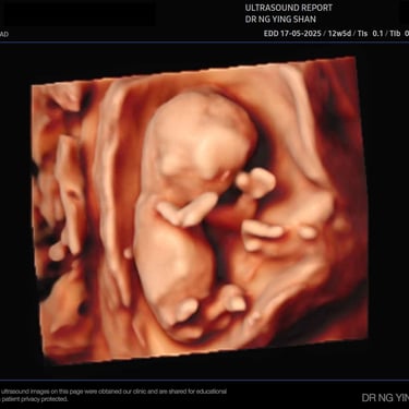
Dating / Early Scan
Confirms the pregnancy is in the uterus, detects the heartbeat, estimates the due date, and determine how many babies
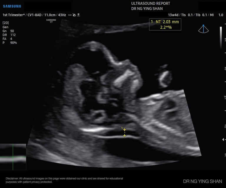
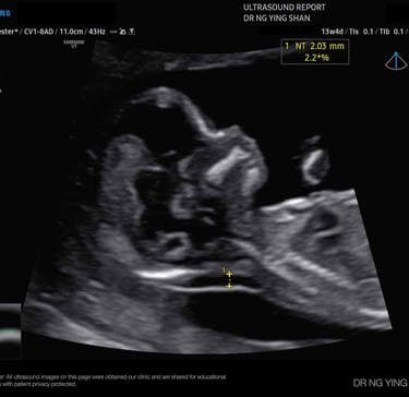
11-14
weeks
First-Trimester Screening
(Nuchal Translucency Scan)
Measures the fluid at the back of the baby’s neck and includes a simple blood test. This helps assess the risk of certain conditions, like Down syndrome, and can also give an early warning for pregnancy complications such as pre-eclampsia.
In simple terms: This scan helps your doctor see if the baby is developing normally and if there are any early concerns.
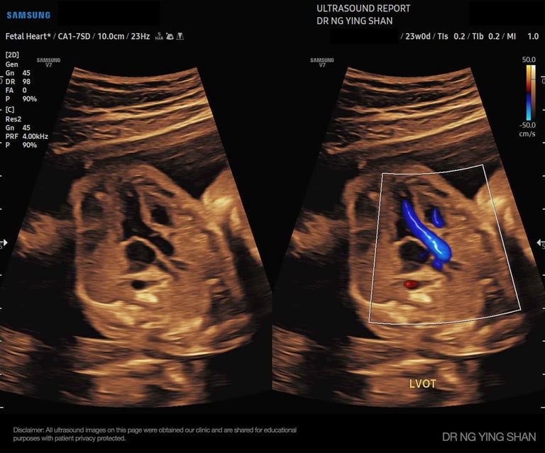
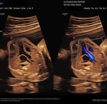
18-22
weeks
Detailed / Anomaly Scan
Looks closely at the baby’s organs, spine, heart, limbs, placenta, and amniotic fluid to check that everything is developing well.
from 24
weeks onwards
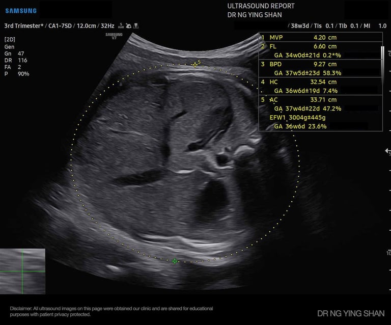
Growth Scans
Monitors the baby’s size, fluid levels, and placenta health, especially if there are concerns about growth or complications.
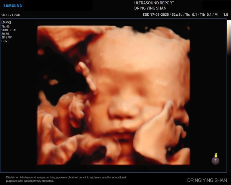
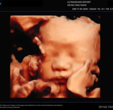
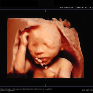
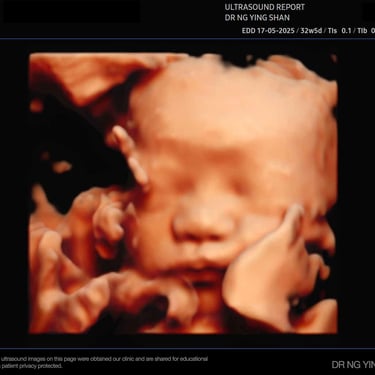
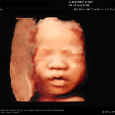
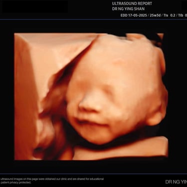
3D/4D Scans (optional)
Provides images of the baby’s face and movements, great for bonding

These scans can detect
Disclaimer: The information provided in this website represents the views and opinions of the original creator. The information provided is intended for informational purposes only and is not intended to be a substitute for professional medical advice, diagnosis, or treatment. It's important to note that this website does not advocate for or endorse any specific tests, products, procedures, opinions, or information mentioned within its content. Always seek the advice of your physician or another qualified health provider for any questions you may have regarding a medical condition.
2026 All rights reserved © Dr Ng Ying Shan
免责声明:此网站提供的信息仅代表原始创作者的观点和意见。此网站提供的信息仅供参考,不旨在替代专业医学建议、诊断或治疗。此网站不主张或支持其内容中提及的任何特定测试、产品、程序、观点或信息。当您遇到任何健康问题时,请咨询您的医生或其他合格的医疗专业人员,,进行诊断与治疗。